- Conserved Hydrogen Bonding Pattern
- Lists of the hydrogen bonds in each of the immunoglobulin variable domains were generated using the program InsightII (MSI/Biosym) and exported as ASCII files. These files were collected in an EXCEL worksheet ans analyzed using the HBond macros.
-
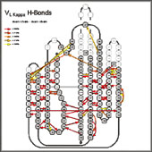
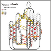
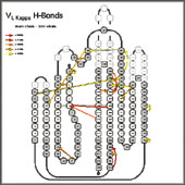
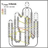
- They were sorted according to domain subtype (VL divided into Vl, Vk and VH into Types I (H6 E, H10 P), Type II (H6 E, H10 G), Type III (H6 Q, H7#P) and Type IV (H6Q, H7P) as described in Honegger & Plückthun, J.Mol.Biol, 309 (2001)687-699).
-
-
|
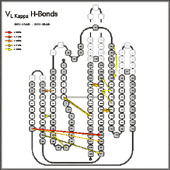
For each pair of sequence positions of each domain subtype, main-chain - main-chain, main-chain - side chain, side-chain - main-chain and side-chain - side-chain hydrogen bonds were counted and the bonds indicated in a schematic representation of the domain, color-coded for the percentage of the domains which actually showed that particular hydrogen bond.
|