Introduction Numbering Sequences Structures Modelling Macros Publications Links
Homology Modelling CDR Grafting Examples HuCAL Biophysical Properties Randomization Catalytic Antibodies

Catalytic Antibodies

Ever since the discovery of catalytic activity in antibodies, researchers have tried to elicit specific catalysts by immunizing animals with transition-state analogs of the reaction to be catalyzed. A number of Fab fragments derived from monoclonal antibodies generated by this procedures, as well as a few engineered Fv fragments, have been successfully crystallized

PDB Files of Catalytic Antibodies:

15C8
1A0Q
1A3L
1A4J
1A4K
1AJ7
1AXS
1C1E
1C5B

1C5C
1CF8
1CT8
1D5B
1D5I
1D6V
1EAP
1F3D
1FIG
1GAF
1HKL
1HYX
1HYY
1KEL
1KEM
1KNO
1MIG
1YEC
1YED
1YEE
1YEF
1YEG
1YEH
1YEI

1YEJ
1YEK
25C8
2RCS
35C8
3FCT
43C9
43CA

(Follow these links to the structures in the PDB database)

Annotated sequence alignments of the VL and the VH domains of these antibodies shows which of the Fabs are related:

Antigen contact analysis reveals which residues are actually involved in Antigen contacts

Binding Modes of Different Antigens

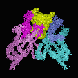
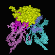
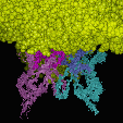
Comparison of the different antibody-antigen complexes in the PDB database shows that the smaller the antigen molecule is, the deeper it has to bury in the antigen combining site to obtain a sufficient ly large interface area: Haptens usually insert into a deep binding pocket, while proteins show a relatively flat binding surface:


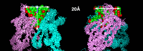
Superposition of Catalytic Antibody Structures

As this superposition of the catalytic antibody structures in the PDB shows, they are typical hapten complexes. The envelope of the superimposed ligands gives an idea of the maximal possible size of the binding pocket: A funnel-shaped cavity of about 20 Å diameter and a depth of about 20 Å, inserted along the axis of the VL/VH heterodimer.

AAAAA Homepage Zürich University Dept. of Biochemistry Plückthun Group Annemarie Honegger

Last Modified by A.Honegger Thursday, November 8, 2001