|
Inhaltsübersicht | Nanomaschinen | Moleküle | Programme | Kurse | Fun | Links |
||
| > |
Dengue Virus
Exploring the Structure
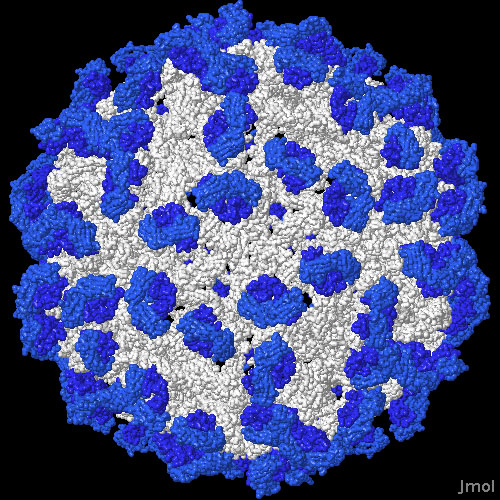
Cryoelectron microscopy has been used to study many aspects of the life cycle of the dengue virus. In these structures, a low resolution image of virus, not quite detailed enough to see atoms, is obtained by the electron microscope, and then atomic structures of the individual pieces are fit into the image to generate the final model. The one shown here, from PDB entry 2r6p, shows the envelope protein on the surface of the virus (in white) with many antibody Fab fragments (in blue) bound to the viral proteins. By looking carefully at this structure, researchers have discovered that the antibodies distort the arrangement of the envelope proteins, blocking their normal action in infection. Other dengue virus structures in the PDB include immature forms of the virus (for instance, in PDB entry 1n6g) and structures that include the membrane-spanning portions of the viral coat (PDB entry 1p58).
This illustration was created with Jmol--you can see an interactive version of the structure by clicking on the image. To see the scientific articles used for this Molecule of the Month and a few questions for further exploration, click here. Also available are related entries in the PDB as determined by a keyword search on June 23, 2008 for dengue virus.
Questions for Further Exploration
|

Previous:
Building
New Viruses
Next: Jmol Animation
Last changed by: A.Honegger,