|
Inhaltsübersicht | Nanomaschinen | Moleküle | Programme | Kurse | Fun | Links |
||
| > |
Cholera Toxin
Exploring the Structure
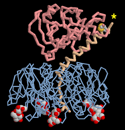
PDB entry 1ltt shows how E. coli enterotoxin finds its target cells in the intestine. The structure includes five molecules of lactose, shown here at the bottom in spacefilling spheres, bound to the targeting portion of the toxin. The carbohydrate chains on a cell surface will bind in the same place when the toxin is attaching to a cell surface. You can also look at how the toxic portion is activated. It is composed of a long, extended portion (colored tan) that anchors the chain to the targeting part. When the toxin is activated, the little loop connecting these parts must clipped and a disulfide linkage must be broken (at the site shown by a star) to release the toxic portion (colored pink) into the cell. In this structure, the little loop is disordered, so the chain looks like it is already broken.This picture was created with RasMol. You can explore these structures by clicking on the accession codes here and picking one of the options under View Structure.
A list of Cholera Toxin related entries in the PDB as determined by a FASTA search on August 25, 2005 is available here. For more information on cholera toxin, click here.
Next: Interaktive 3D-Animation
Previous: Terrible Toxins
Last changed by: A.Honegger,
