|
Inhaltsübersicht | Nanomaschinen | Moleküle | Programme | Kurse | Fun | Links |
||
| > |
Major Histocompatibility Complex

Exploring the Structure
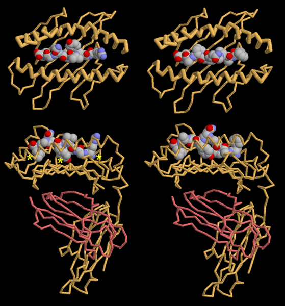
The entire MHC system poses a problem: each cell has thousands of different peptides to display, but each cell only builds a few types of MHC. The solution to this dilemma was revealed in the early structures of MHC with different peptides. The two structures shown here, entry 2vaa on the left and entry 2vab on the right, have peptides from two different viruses bound to the same MHC. Another similar series can be found in PDB entries 1hhg, 1hhh, 1hhi, 1hhj, and 1hhk. Looking at these structures, you can see that the peptide, which is nine amino acids long, is held in an extended conformation in a groove between two long alpha helices, as shown in the upper pictures. The MHC protein grips the peptide at each end and at a tyrosine in the middle, as shown by the yellow stars. Notice that these three positions are similar in the two structures. The peptide is anchored to MHC at these points, but the other amino acids extend outward, away from the protein. Next month, we will look at how the immune system recognizes these exposed portions of the peptides.These illustrations were created with RasMol. You can create similar pictures by clicking on the accession codes here and picking one of the options under View Structure.
 A list of all MHC entries in the PDB as determined by a FASTA search on January 26, 2005 is available here. For additional reading on MHC, click here.
Next: Interaktive 3D-Animation
Previous: A Family of Folds
Last changed by: A.Honegger,