|
Inhaltsübersicht | Nanomaschinen | Moleküle | Programme | Kurse | Fun | Links |
||
| > |
The Glycolytic Enzymes

Fructose 1,6-bisphosphate Aldolase
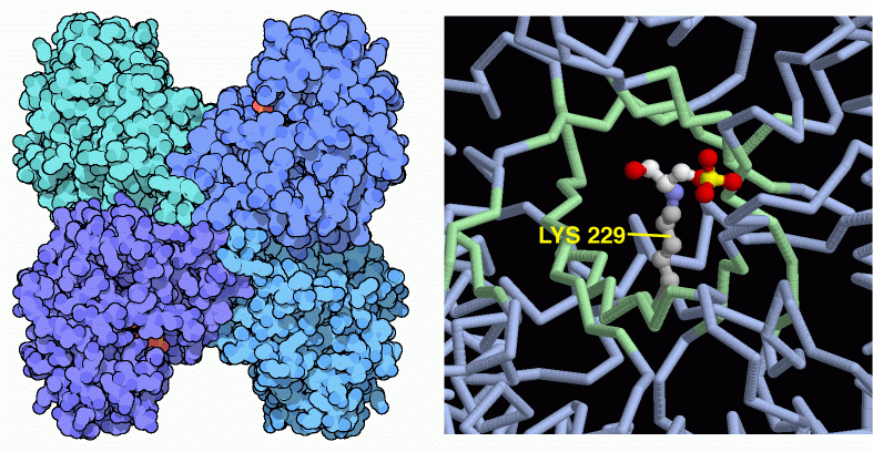
At this stage in glycolysis, the sugar molecule is primed and the cell is ready to start breaking it up. The fourth enzyme, fructose 1,6-bisphosphate aldolase, cuts the molecule in the middle, producing two similar pieces, each with a single phosphate attached. The enzyme also readily performs the reverse reaction, connecting these two smaller molecules to reform the phosphorylated fructose. In fact, the enzyme is named for this reverse reaction, which is an aldol condensation. The enzyme shown here, from PDB entry 4ald, is found in our muscle cells. It contains four identical subunits, each with its own active site. The active site uses a special lysine, number 229 in this particular form, to attack the sugar chain. As shown on the right in PDB entry 1j4e, this lysine forms a covalent bond with molecule during the cleavage reaction. This structure is frozen at a stage when the sugar molecule (with red oxygen, white carbon, and yellow phosphorus) has been cleaved and only half is left in the active site.The aldolase enzyme used by most bacteria is different than the aldolases that we have in our cells. It uses two magnesium ions instead of a special lysine amino acid. You can look at an example of a bacterial aldolase in PDB entry 1zen. Be sure to look for the metal ions! Also, take a look at the unusual enzyme made by hot-spring archaebacteria, in PDB entry 1ojx. It uses a lysine in the active site, like our enzyme, but is composed of ten chains in a huge molecular complex.
Next: 5: Triose Phosphate Isomerase
Previous: 3: Phosphofructokinase
Last changed by: A.Honegger,