|
Inhaltsübersicht | Nanomaschinen | Moleküle | Programme | Kurse | Fun | Links |
||
| > |
Calmodulin
Exploring the Structure
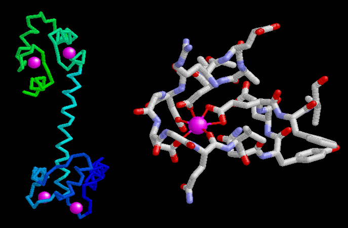
Calmodulin contains four nearly identical high-affinity calcium binding sites, as seen in the backbone diagram of PDB entry 1cll shown on the left. The calcium ions are shown in purple. The calcium-binding motif is comprised of a characteristic loop flanked by two alpha helices. As shown on the right, the positively-charged calcium ion is surrounded in the loop by negatively-charged sidechains of three aspartates and one glutamate, as well as one oxygen atom from the backbone of the protein chain.This illustration was created using RasMol. You can create similar illustrations by clicking on the accession code above and then picking one of the options under View Structure.
A list of all calmodulin structures in the PDB as of August, 2003, is available here. For more information on calmodulin, click here. To look at calmodulin from the perspective of genomics, take a look at the Protein of the Month feature at the European Bioinformatics Institute.
Next: Interaktive 3D-Animation
Previous: Flexibility of Calmodulin

Last changed by: A.Honegger,