|
Inhaltsübersicht | Nanomaschinen | Moleküle | Programme | Kurse | Fun | Links |
||
| > |
Src Tyrosine Kinase
Exploring the Structure
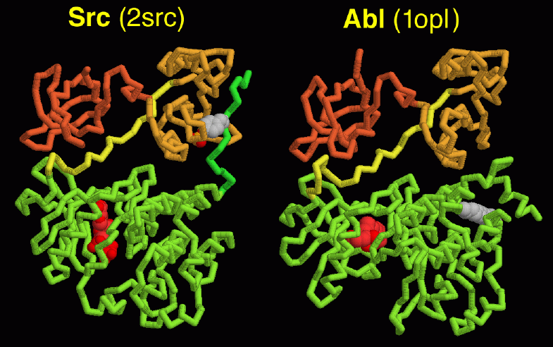
When exploring oncogene proteins, PDB entry 2src is a good place to start. This is the inactive form of Src, folded into a tight ball. The structure has a nucleotide bound in the kinase active site (shown in red here), and the key tyrosine (number 527 in the chain, seen here on the right side of the molecule) has a phosphate attached. You can also look at the Hck protein, PDB entry 2hck, which is almost identical to Src. A fragment of the Abl protein, which at full-length is much larger than Src and Hck, is also available in PDB entry 1opl. It is slightly different than Src and Hck, and doesn't use a tail to stabilize the closed form. This structure includes a drug molecule in the active site (shown in red here) and a lipid bound in a deep pocket (shown in gray).These images were created with RasMol. You can create similar images by clicking on the accession code above and then choosing one of the options under View Structure. Note that the 2hck and 1opl structures each have two separate chains: you will want to look at only chain A in each case.
For additional information on Src protein, click here.

Next: Interaktive 3D-Animation
Previous: Many Moving Parts
Last changed by: A.Honegger,