|
Inhaltsübersicht | Nanomaschinen | Moleküle | Programme | Kurse | Fun | Links |
||
| > |
Thrombin
Exploring the Structure
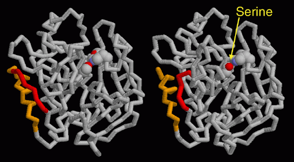
PDB entry 1mkx is a perfect structure for exploring thrombin. It contains two molecules of the protein, one in inactive form (chain K) and one activated (chain H and L). The inactive form is shown on the left. In order to activate the protein, the protein strand must be cleaved between the yellow and red segments on the left side. Then, the two new ends separate and the whole protein relaxes into the active form, shown on the right. Notice how, in the active form, the key catalytic serine amino acid, with oxygen atom in bright red, changes position and points straight out into the active site, ready to perform the cleavage.This picture was created with RasMol. You can create similar pictures by clicking on the accession codes above and choosing one of the options under View Structure.
A list of all thrombin structures in the PDB as of January, 2002, is available here. For additional information on thrombin, click here.
Next: Interaktive 3D-Animation
Previous: Anticoagulants
Last changed by: A.Honegger,
