|
Inhaltsübersicht | Nanomaschinen | Moleküle | Programme | Kurse | Fun | Links |
||
| > |
Glycogen Phosphorylase
Although it may not seem so during the holiday season, we do not have to eat continually throughout the day. Our cells do require a constant supply of sugars and other nourishment, but fortunately our bodies contain a mechanism for storing sugar during meals and then metering it out for the rest of the day. The sugars are stored in glycogen, a large molecule that contains up to 10,000 glucose molecules connected in a dense ball of branching chains. Your muscles store enough glycogen to power your daily activities, and your liver stores enough to feed your nervous system and other tissues all through the day and on through the night.
Sweet Tooth
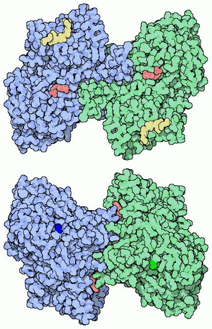
Sugar is released from glycogen by the enzyme glycogen phosphorylase. It clips glucose from the chains on the surface of a glycogen granule. The enzyme is a dimer of two identical subunits (colored green and blue in the structure here, from PDB entry 6gpb). In the upper illustration, two nucleotides, in red, are bound at the entrance to the active site, which is found in a deep cleft. The yellow molecules are short chains of sugars similar to the ends of glycogen chains, which bind into another cleft that the enzyme uses to grip the glycogen granule. In its cleavage reaction, glycogen phosphorylase uses a phosphate molecule, connecting it to the sugar as it is released. A second enzyme, phosphoglucomutase, then shifts the position of the phosphate to a neighboring carbon atom in the sugar, making the sugar ready for breakdown by glycolysis.Moderation
As you might imagine, this process is highly regulated. Traffic of sugar into and out of storage in glycogen is used to control the level of glucose in the blood, so glycogen phosphorylase must be activated when sugar is needed and quickly shut down when sugar is plentiful. It is controlled in several ways. First, the enzyme is activated by adding a phosphate molecule to a serine amino acid (serine 14) on the back side of the enzyme, shown in bright green and blue in the lower illustration. The phosphate causes a large shift in the shape of the enzyme (shown on the next page), shifting it into the active conformation. Two special enzymes control the addition and removal of this phosphate, based on levels of the sugar-monitoring hormones insulin and glucagon, and other hormones such as epinephrine (adrenaline).Also, binding of other molecules can modify the activity of the molecule. For instance, AMP (adenine monophosphate) binds to a different site on the back side of the molecule (shown in red on the lower illustration), causing the same shift to the active conformation. This is useful, because AMP is a product of ATP breakdown and will be more plentiful when energy levels are low and more sugar is needed.
Next: Shape-shifting
Last changed by: A.Honegger,
