|
Inhaltsübersicht | Nanomaschinen | Moleküle | Programme | Kurse | Fun | Links |
||
| > |
Myosin
Exploring the Structure
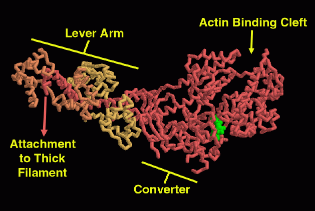
This myosin motor domain, from entry 1b7t, is nearly straight, close to the rigor form. You can explore several interesting features. At the tip of the molecule is a cleft that binds to the actin filament. Notice that the ADP molecule (in green) is bound at the base of this deep cleft. It is thought that changes in the nucleotide, as it cycles from ATP, to ADP and phosphate, to ADP alone, are transmitted along this cleft to change the way that myosin interacts with actin. In the middle of the molecule is the "converter" domain that changes shape when phosphate is released. On the left side of the molecule is a long alpha helix with the two light chains (orange and yellow) bound around it. This is the "lever arm" that amplifies the converter shape change into a large power stroke.This illustration was created using RasMol, using tubes to show the protein backbones. You can create similar pictures by clicking on the PDB accession code above and picking one of the options under "View Structure."
A list of all myosin structures as of June, 2001, is available here. For suggestions for further reading about myosin, click here.
Next: Interaktive 3D-Animation
Previous: The Power Stroke

Last changed by: A.Honegger,