|
Inhaltsübersicht | Nanomaschinen | Moleküle | Programme | Kurse | Fun | Links |
||
| > |
Myoglobin
Oxygen Bound to Myoglobin
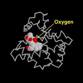
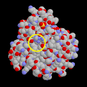
A later structure of myoglobin, with the accession code 1mbo, shows the location of oxygen. The iron atom at the center of the heme group holds the oxygen molecule tightly. Compare the two pictures. The first shows only a set of thin tubes to represent the protein chain, and the oxygen is easily seen. But when all of the atoms in the protein are shown in the second picture, the oxygen disappears, buried inside the protein.
So how does the oxygen get in and out, if it is totally surrounded by protein? In reality, myoglobin (and all other proteins) are constantly in motion, performing small flexing and breathing motions. Temporary openings constantly appear and disappear, allowing oxygen in and out. The structure in the PDB is merely one snapshot of the protein, caught when it is in a tightly-closed form. Looking at the static structure held in the PDB, we must imagine the dynamic structure that actually exists in nature.
The two pictures above were created with RASMOL. You can create similar pictures by accessing the PDB file 1mbo, and then clicking on "View Structure." Try switching between the two types of pictures shown above, to prove to yourself that the oxygen is buried in this structure!
Next: Interaktive 3D-Animation
Previous: A Closer Look


Last changed by: A.Honegger,