|
Inhaltsübersicht | Nanomaschinen | Moleküle | Programme | Kurse | Fun | Links |
||
| > |
Glucose Oxidase

Exploring the Structure
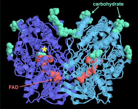
You can take a closer look at glucose oxidase from a Penicillium mold in PDB entry 1gpe. The oxidation reaction is performed by the FAD cofactors bound deep inside the enzyme, shown in red. The active site where glucose binds is just above the FAD, in a deep pocket shown with a star. Notice that the enzyme, like many proteins that act outside of cells, is covered with carbohydrate chains, shown in green.
This picture was created with RasMol. You can create similar pictures by clicking on the accession codes here and picking one of the options under Images and Visualization.
A list of 'glucose oxidase' related entries in the PDB as determined by a keyword search on April 28, 2006 is available. For additional reading on glucose oxidase, click here.
Next: Interaktive 3D-Animation
Previous: Choices for Biosensors
Last changed by: A.Honegger,