|
Inhaltsübersicht | Nanomaschinen | Moleküle | Programme | Kurse | Fun | Links |
||
| > |
Acetylcholine Receptor

Exploring the Structure
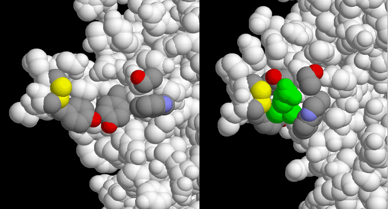
Although there aren't currently structures for both the open and closed states of the acetylcholine receptor, you can see what happens when acetylcholine binds by looking at a similar protein, acetylcholine-binding protein. This protein was discovered in certain sea slugs, where it modulates the signals carried by acetylcholine. It is very similar to the outer part of the acetylcholine receptor, without the membrane-crossing part. The binding site of the acetylcholine receptor (PDB entry 2bg9) is shown here on the left, in the closed state before acetylcholine binds. The important amino acids in the binding site are shown in atomic colors, including an unusual disulfide linkage between two adjacent cysteines. The similar portion of the acetylcholine binding protein (PDB entry 1uv6) is shown on the right, with acetylcholine bound (shown in green). Notice that these amino acids fold up around the neurotransmitter. As the binding site closes around acetylcholine, it shifts the conformation of the whole receptor, opening the pore through the membrane.These pictures were created with RasMol. You can create similar pictures by clicking on the accession codes here and picking one of the options under View Structure. The amino acids highlighted in acetylcholine receptor are Trp149, Thr150, Tyr190, Cys192, Cys193, and Tyr198 of chain A. The similar amino acids in acetylcholine-binding protein are Trp143, Thr144, Tyr185, Cys187, Cys188, and Tyr192.
A list of Acetylcholine Receptor related entries in the PDB as determined by a FASTA search on October 27, 2005 is available here. For more information on the acetylcholine receptor, click here.
Next: Interaktive 3D-Animation
Previous: Cobras and Curare
Last changed by: A.Honegger,