|
Inhaltsübersicht | Nanomaschinen | Moleküle | Programme | Kurse | Fun | Links |
||
| > |
Carotenoid Oxygenase

Exploring the Structure
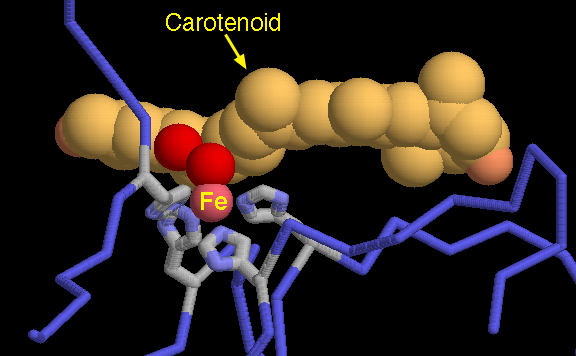
PDB entry 2biw is the first structure of a carotenoid oxygenase, revealing the mechanistic details of this important class of enzymes. The picture shown here is a close-up of the active site. The carotenoid, shown in orange, is held close to an iron atom, in pink. Two water molecules, shown here with red spheres, are thought to be positioned approximately where the oxygen will be bound when the cleavage reaction is performed. Notice that the iron atom is gripped by four perfectly-positioned histidine amino acids.This picture was created using RasMol. You can create similar illustrations by clicking on the accession codes here and picking one of the options under View Structure. When you go to look at this structure, notice that the PDB file contains four separate molecules of the enzyme. You will probably want to look at just one of the chains.
A list of carotenoid oxygenase entries in the PDB as determined by a FASTA search on June 02, 2005 is available here. For more information on carotenoid oxygenases, click here.
Next: Interaktive 3D-Animation
Previous: Carotene and Retinal in Action
Last changed by: A.Honegger,