|
Inhaltsübersicht | Nanomaschinen | Moleküle | Programme | Kurse | Fun | Links |
||
| > |
Cytochrome C

Exploring the Structure
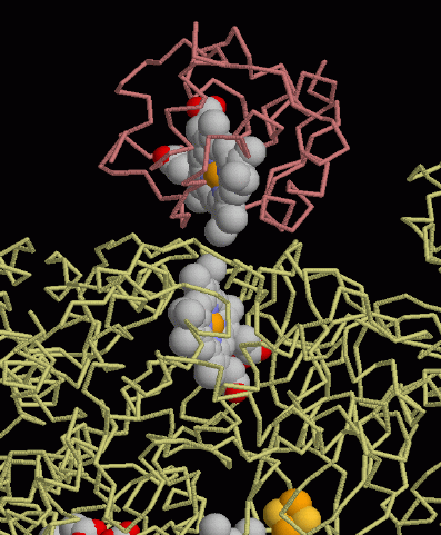
PDB entry 1kyo gives a close-up view of how electrons are transferred between carriers inside cells. Electrons do not flow through a continuous wire, like in our familiar appliances. Instead, at these small distances electrons tunnel directly from one carrier to the next. This picture shows the complex of cytochrome c (at the top) and the large cytochrome bc1 complex, shown at the bottom. The protein chain in cytochrome c is shown in pink tubes and the protein chains in the bc1 complex are shown in yellow tubes. The hemes are shown with spheres at each atom, with the iron atoms in yellow. Notice how the heme group of cytochrome c is pushed close to a heme group in the bc1 complex. At this distance, electrons tunnel from one heme to the next in less than a millionth of a second.This picture was created with RasMol. You can create similar illustrations by clicking on the accession code above and picking an option under View Structure.
A list of all cytochrome c structures in the PDB as of December, 2002, is available here. For more information on cytochrome c, click here.
Next: Interaktive 3D-Animation
Previous: Cellular Circuits
Last changed by: A.Honegger,