|
Inhaltsübersicht | Nanomaschinen | Moleküle | Programme | Kurse | Fun | Links |
||
| > |
Ferritin and Transferrin
Exploring the Structure
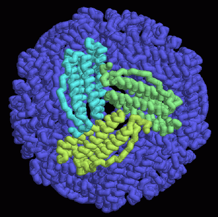
The way that iron atoms get into and out of ferritin is still something of a mystery. Many researchers believe that they enter through the small pores between the 24 subunits. A few of the pores at four-fold axes are shown on the first page, and the pore at one of the three-fold axes is shown in the middle here, from PDB entry 1mfr. Once inside, iron atoms form crystallites that are too large to get back out of the pores. So, in order to release the iron atoms, the entire complex must be disassembled.Here is a hint for when you look at these structures yourself. The PDB entry 1mfr (shown here) has all 24 chains in the file, but if you want to look at other structures, such as human forms and horse forms, the PDB file only contains one subunit. To get coordinates for the whole complex, use the link to the EBI MSD Macromolecule File Server under the "Other Sources" section of the Structure Explorer page for this entry. Then, you can create pictures similar to this one, using programs such as RasMol or the other programs found under "View Structure."
A list of all dihydrofolate ferritin and transferrin in the PDB as of November, 2002, is available here. For more information on ferritin and transferrin, click here.
Next: Interaktive 3D-Animation
Previous: Transporting Iron Ions
Last changed by: A.Honegger,
