|
Inhaltsübersicht | Nanomaschinen | Moleküle | Programme | Kurse | Fun | Links |
||
| > |
Bacteriorhodopsin
Exploring the Structure
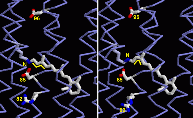
Many structures of bacteriorhodopsin are available in the PDB, showing many of the steps in the process of absorbing light and pumping protons. Two snapshots are shown here. The structure on the left, from PDB entry 1c3w, is in the ground state, before it has absorbed light. The retinal (the long molecule in white crossing through the center) is in the straight trans form, as shown by the little zig-zag yellow lines. The structure on the right, from PDB entry 1dze, shows the molecule after absorbing light. Notice that the retinal now has a bent cis shape at the site indicated by the yellow lines. This new shape has changed the orientation of the nitrogen at the end of the retinal, indicated by the yellow N. It has also shifted the position of several protein amino acids that are along the pathway of proton transfer, indicated by the numbers. In particular, notice the large shift of arginine 82 at the bottom. Researchers are working to discover how these changes in shape power the transfer of a proton from the top to the bottom, through the middle of bacteriorhodopsin and across the archaebacterial membrane.This picture was created with RasMol. You can create similar pictures by clicking on the accession codes above and picking one of the options under View Structure.

A list of all bacteriorhodopsin structures in the PDB as of March, 2002 is available here. For suggestions for additional reading about bacteriorhodopsin, click here.
Next: Interaktive 3D-Animation
Previous: Seeing Light
Last changed by: A.Honegger,