|
Inhaltsübersicht | Nanomaschinen | Moleküle | Programme | Kurse | Fun | Links |
||
| > |
Poliovirus and Rhinovirus

Picornavirus Structure
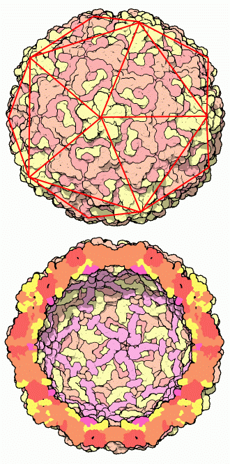
Many viruses, including the picornaviruses and bacteriophage phiX174 (discussed in an earlier Molecule of the Month), are icosahedral in shape. They are composed of 60 identical pieces that form a perfectly symmetrical shell, termed a capsid, around the viral genome. In the case of poliovirus and rhinovirus, the shell is composed of 60 copies of four different proteins (colored yellow, orange, red, and magenta on rhinovirus here, PDB entry 4rhv), for a total of 240 protein chains in all. Notice that the fourth chain, colored magenta, can only be seen on the inside surface of the capsid. These proteins are carefully designed to be stable, but not too stable. They must be fairly sturdy to allow the virus to pass from host to host through the hostile environment. But at the same time, they must be able to fall apart when they enter the cell, releasing the RNA inside. A carefully orchestrated set of structural changes occur as the virus attaches to the surface of the cell and is drawn inside, allowing the virus to deliver its RNA into the unwitting host.The RNA protected inside the capsid is seen only as a blurry tangle in these crystallographic structures and is not shown in these pictures, because it is not as perfectly symmetrical as the many proteins in the shell. The rhinovirus genome, when analyzed by sequencing techniques, contains just enough information to direct the construction of eleven proteins. These include the four separate proteins for its capsid, another four proteins that replicate its RNA, two proteins to clip each of these proteins into the proper shape, and one additional protein with as-yet obscure function.
Next: Antibody Protection
Previous: Poliovirus and Rhinovirus
Last changed by: A.Honegger,