|
Inhaltsübersicht | Nanomaschinen | Moleküle | Programme | Kurse | Fun | Links |
||
| > |
Actin
Exploring the Structure
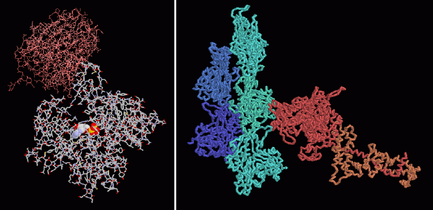
Large helical protein assemblies, such as actin filaments, are notoriously difficult to study by crystallography, because the filaments do not form perfect crystals. The structures of actin in the PDB all have something bound to them, blocking formation of a filament, so the structures contain only a single actin molecule, not an entire actin filament. PDB entry 1atn, shown at the left, contains a DNA-cutting enzyme (colored pink) that just happens to bind to actin. Actin is a U-shaped molecule with ATP (shown in spacefilling spheres) bound deep in the groove between the two arms. PDB entry 1alm, shown on the right, presents a model of one myosin motor (red and yellow) bound to a short actin filament formed of five molecules (blue), based on data from electron microscopy. The file contains only alpha carbon positions for the proteins, so you'll need to use backbone diagrams like the one shown here when you look at it.These illustrations were created with RasMol. To create similar illustrations, click on the PDB accession codes above and pick one of the options under View Structure. A list of all actin structures in the PDB as of July, 2001, is available here. For more information about actin, click here.
Next: Interaktive 3D-Animation
Previous: Controlled Growth
Last changed by: A.Honegger,
