|
Inhaltsübersicht | Nanomaschinen | Moleküle | Programme | Kurse | Fun | Links |
||
| > |
Insulin
Exploring the Structure
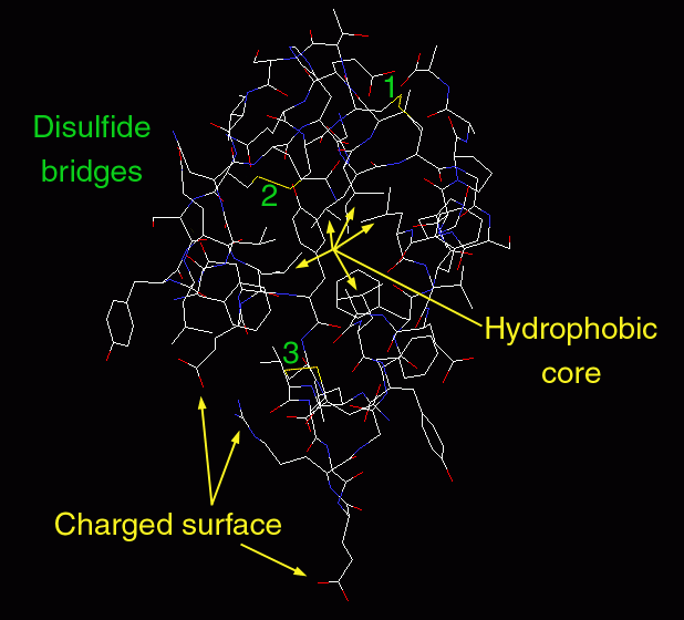
Insulin is a perfect molecule for exploring protein structure. It is small enough that you can display all of the atoms and still have a picture that is not too confusing. Human insulin is pictured here, using entry 1trz. The file contains four chains, labeled A, B, C, and D. When looking at this structure yourself, you will want to display only the A and B chains, which together compose one monomer of insulin. In the structure, you can see many of the key features that stabilize protein structure. Notice the cluster of carbon-rich amino acids, like leucine and isoleucine, that cluster in the middle of insulin, forming a hydrophobic core. Notice that the surface is covered with the charged amino acids lysine, arginine, and glutamate. These amino acids interact favorably with the surrounding water. Also notice the three disulfide bridges between cysteine amino acids, which stabilize this tiny protein.
This picture was created with RasMol. You can create similar images by clicking on the accession code above, and choosing one of the molecular viewers available through the link "View Structure."
Next: Interaktive 3D-Animation
Previous: Insulin Therapy

Last changed by: A.Honegger,