|
Inhaltsübersicht | Nanomaschinen | Moleküle | Programme | Kurse | Fun | Links |
||
| > |
Restriction Enzymes

Exploring the Structure
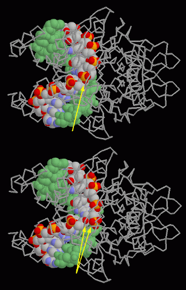
The PDB contains structures for many restriction enzymes. Another example from Escherichia coli--EcoRV--is shown here. The structure at the top, taken from PDB entry 1rva, shows the enzyme bound to a short piece of DNA. The arrow shows the phosphate group that will be cut. The lower illustration, taken from PDB entry 1rvc, shows the structure after the DNA has been cut. A water molecule has been inserted, so there are now two oxygen atoms, close to one another but not bonded together, where there was a single bonded oxygen atom in the intact DNA.
In both illustrations, the protein is shown with a simple backbone representation and one DNA strand is colored green. These illustrations were created with RasMol. You can create similar illustrations by clicking on the PDB accession codes above, and then picking "View Structure".
Next: Interaktive 3D-Animation
Previous: Sticky Ends
Last changed by: A.Honegger,