|
Inhaltsübersicht | Nanomaschinen | Moleküle | Programme | Kurse | Fun | Links |
||
| > |
Bacteriophage phiX174
Exploring the Structure
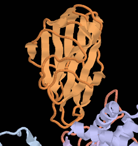
You can easily look at one of the subunits of this bacteriophage. There are seven separate chains in the PDB file 1cd3. The spike protein, chain G, is small and compact and the capsid protein, chain F, is large. Both are very stable structures composed of two beta-sheets, forming a structure commonly called a "beta-sandwich." The ribbon diagram shows the chain of the spike protein. Notice how the beta-strands, each depicted with an arrow, arrange side-by-side to form the two sheets. Beta-sandwich structures like this are found in many different viruses.
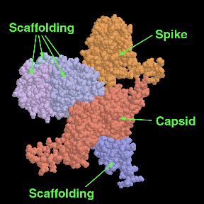
Four copies of a small scaffolding protein (chains 1, 2, 3 and 4) are arranged in the angle between the capsid and spike proteins, ending up on the outside of the final virus capsid. Another small scaffold protein (chain B) is found on the inside of the capsid, where it assists in the capture of DNA. Compare this detailed atomic view of one subunit of the capsid to the picture of the whole capsid shown on the previous pages, which contains 60 identical copies of each of these seven proteins.
The pictures were created with RasMol. You can create similar pictures by accessing the PDB file 1cd3 and then clicking "View Structure."
Next: Interaktive 3D-Animation
Previous: Assembling a Virus


Last changed by: A.Honegger,