|
Inhaltsübersicht | Nanomaschinen | Moleküle | Programme | Kurse | Fun | Links |
||
| > |
Cadherin
Exploring the Structure
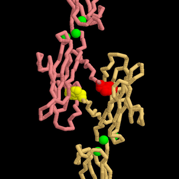
The adhesive contact between two cadherin proteins is formed when a tryptophan on one protein extends over and binds in a pocket on the other protein. The contact is shown here from PDB entry 1l3w. Notice the three calcium ions (in green) between the domains, which rigidify the whole structure. If you want to explore this interaction yourself, PDB entry 1l3w is a tricky structure to use, since you have to perform a crystallographic transformation to get the second protein structure (the transformation is a 180 degree rotation around the Y axis, which may be obtained with coordinates -x, y, -z). You can easily see the interaction, though, by looking at PDB entry 1nci, which only includes the last domain in each cadherin chain, but has two proteins in the PDB file.
These pictures were created with RasMol. To create similar pictures, you can click on the accession codes here and pick one of the options under Images and Visualization. To see the scientific papers used to research this Molecule of the Month, click here. Also available are related entries in the PDB as determined by a keyword search on February 27, 2008 for cadherin.
Previous: Gluing Cells Together

Last changed by: A.Honegger,