|
Inhaltsübersicht | Nanomaschinen | Moleküle | Programme | Kurse | Fun | Links |
||
| > |
Serpins
Exploring the Structure
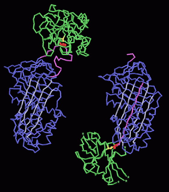
These two amazing crystallographic structures show before and after pictures of alpha1-antitrypsin action. PDB entry 1k9o, on the left, shows the serpin-protease complex before it is trapped. The scientists used a mutant form of the protease, with the reactive serine changed to an alanine (shown in yellow), to look at the complex without tripping the trap. Notice the four long parallel strands in the serpin, colored white. PDB entry 1ezx, on the right, shows the complex after it has snapped. The loop has broken, and the trypsin has been dragged all the way down to the other side of the serpin. The trypsin serine (yellow) is attached with a bond to the serpin methionine (red). Notice how the strand from the broken loop has zipped into the middle of the four parallel strands. Notice also that the trypsin is destabilized. Much of the protein chain was not seen in the crystal structure because it was moving too much. The little stars show places where the experimental structure ends and loops are not seen.This picture was created using RasMol. You can create similar pictures by clicking on the accession codes and picking one of the options under View Structure.
A list of all PDB entries related to Serpins as of April, 2004 is available here. For more information on serpins, click here.
Next: Interaktive 3D-Animation
Previous: Tripping the Trap
Last changed by: A.Honegger,
