|
Inhaltsübersicht | Nanomaschinen | Moleküle | Programme | Kurse | Fun | Links |
||
| > |
The Calcium Pump

Exploring the Structure
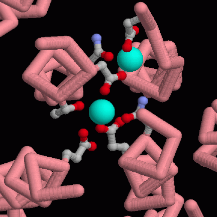
The calcium binding site is in a tunnel formed by four alpha helices, which cross straight through the membrane. This illustration, from PDB entry 1eul, shows a view down the helices. The two calcium ions, shown as blue-green spheres, are held by a collection of amino acids, shown in balls-and-sticks, that coordinate it from all sides. The protein is far less stable when these calcium ions are removed. You can look at the structure of the calcium-free form in PDB entry 1iwo. It was solved by adding a drug molecule that binds near the calcium-binding site and freezes the protein into a stable, but non functioning, form.This picture was created with RasMol. You can create similar pictures by clicking on the PDB accession codes and picking one of the options under View Structure.
A list of all PDB entries related to Calcium Pumps as of February, 2004 is available here. For more information on calcium pumps, click here.
Next: Interaktive 3D-Animation
Previous: The Pumping Cycle
Last changed by: A.Honegger,