|
Inhaltsübersicht | Nanomaschinen | Moleküle | Programme | Kurse | Fun | Links |
||
| > |
Rubisco (Ribulose Bisphosphate Carboxylase/Oxygenase)
Exploring the Structure
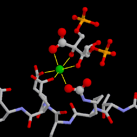
The active site of rubisco is arranged around a magnesium ion. In this picture, drawn using coordinates from PDB entry 8ruc, the magnesium ion is shown at the center in green. Above it is a small sugar molecule that is similar to the product of the rubisco reaction, and a short stretch of the protein chain is shown at the bottom. In reality, the rubisco protein chains completely surround these molecules but are not shown here for clarity.
The magnesium ion is held tightly by three amino acids, including a surprising modified form of lysine (the bonds between the ion and the protein are shown by the three yellow lines going downwards). An extra carbon dioxide molecule, shown in larger spheres just below the magnesium ion, is attached firmly to the end of the snaky lysine sidechain. In plant cells, this "activator" carbon dioxide, which is different from the carbon dioxide molecules that are fixed in the reaction, is attached to rubisco during the day, turning the enzyme "on," and removed at night, turning the enzyme "off." The exposed side of the magnesium ion is then free to bind to both ribulose bisphosphate, holding onto two oxygen atoms (small red spheres), and the carbon dioxide molecule that will be attached to sugar. In this structure, the carbon dioxide, shown with larger spheres above the magnesium ion, is already attached to the sugar.
This illustration was created with RasMol. You can create similar illustrations by going to entry 8ruc and clicking "View Structure." You will find that this structure includes only one half of the entire rubisco complex--if you are interested in looking at the whole rubisco molecule, the structure in 1rcx contains all sixteen chains.
Next: Interaktive 3D-Animation
Previous: Sixteen Chains in One
Last changed by: A.Honegger,
