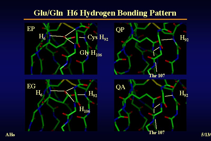
 Hydrogen Bonding Patterns of H6 Glu and H6 GlnThe two different hydrophilic side chains show distinct hydrogen bonding patterns, but both average close to four hydrogen bonds over all structures in the pdb database,except in those structures in which the O and N of the glutamine H6 side chain have been swapped. (Gln makes an additional H-bond (not drawn) to the side chain of Gln H105) If one compares the structure of different members of the immunoglobulin superfamily, it becomes apparent that the presence of Glu or Gln in position 6 correlates both with the kink in the chain in pos. 8-10 and the beta-bulge of the gly-X-gly motive in posion H104-H106. It is well possible that these conformational peculiarities of the variable domain subclass of the immunoglobulin superfamily represent an adaptation to the need to provide hydrogen bonding partners for a buried polar side-chain. |